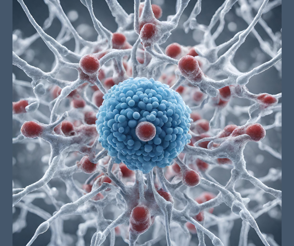
The cell cytoskeleton is modulated by motor proteins, known as dyneins, that generate force and movement on microtubules to control a wide range of biological processes, including motility, cell division, and intracellular transport.
Mutation of Lis1, a key dynein regulator, can lead to the rare mental developmental disorder lissencephaly, or “smooth brain,” in which the brain’s surface lacks normal folds and grooves. There is currently no cure.
In a new study published in Nature Structural and Molecular Biology, titled “Multiple steps of dynein activation by Lis1 visualized by cryo-EM,” researchers from the Salk Institute and UC San Diego (UCSD) catalogued new high-resolution dynamics of Lis1 and dynein to guide the design of drugs targeting neurological diseases. Using a yeast model, the team solved 16 new structural conformations of the dynein–Lis1 complex using cryo-electron microscopy (cryo-EM) to pinpoint atomic locations critical for drug targeting.
“Our approach to imaging dynein and Lis1 is more comprehensive than any previous study on this protein,” said co-corresponding author Andres Leschziner, PhD, professor at UCSD. “By capturing movies rather than pictures, we confirmed 16 detailed, 3D shapes that dynein takes as it interacts with Lis1, several of which are entirely unique to our study.”
Dynein is part of the AAA+ ATPase family, using energy from ATP hydrolysis to generate motion. In cells, dynein typically remains in an autoinhibited “Phi” or “locked” conformation, detached from microtubules. Lis1 plays a crucial role in switching dynein into its active “unlocked” state and helping it assemble into complexes. Evidence shows that dynein mutants in the unbound state bind Lis1 more strongly, suggesting that Lis1 acts before dynein interacts with microtubules. However, the detailed structural mechanism remained unclear until now.
Unlike previous studies that provided snapshots of dynein’s locked or unlocked forms, the Salk and UCSD team created a time-resolved “movie” capturing the sequence of structural transitions that occur as dynein activates—documenting the sub-second changes involved in this process.
The dynein complex consists of a dimer of two motor domains and two copies of five accessory chains (intermediate chain, light intermediate chain, and three light chains). The heavy chain motor domain is composed of six AAA+ modules, each with a large and small domain, with nucleotide binding occurring between adjacent modules.
The study suggests that Lis1 relieves dynein autoinhibition by increasing its basal ATP hydrolysis rate, encouraging conformations that enable complex assembly and motility. The data support a model where Lis1 facilitates nucleotide exchange at AAA3, potentially priming dynein for assembly and activation.
“The impressive tools we have today in the lab made it possible for us to create a movie of dynein and Lis1 interacting in real time,” said Agnieszka Kendrick, PhD, assistant professor at Salk and co-corresponding author. “Having this detailed view of their stepwise partnership will help us find ways to restore their activity in neurodevelopmental and neurodegenerative diseases.”
These new high-resolution 3D insights into dynein–Lis1 interactions could pave the way for treating dysfunction in related brain disorders. Future studies aim to examine how various Lis1 mutations impact these interactions and contribute to lissencephaly and other rare genetic diseases.
Source: Genetic Engineering & Biotechnology News, May 26, 2025