By Quan-Sheng Shu, Ray Radebaugh, and Robert L. Fagaly
Cryogenics and superconductivity (SC) have transformed modern medicine. From noninvasive tumor treatments to the imaging power of MRI and SQUID-based diagnostics, these technologies have become essential tools across the biomedical landscape. This article traces key developments over the past 50 years and highlights how cryogenics and superconductivity are shaping the next generation of diagnostics and therapy.
Cryoablation, a technique that uses extremely low temperatures to destroy abnormal tissue, has emerged as one of the most important advances. Originally demonstrated in 1961 with vacuum-insulated cryoprobes cooled by liquid nitrogen, cryoablation now treats conditions ranging from cancer to cardiac arrhythmias. Minimally invasive techniques—like catheter-based systems or those introduced transbronchially—allow access to internal organs without open surgery. Ultrasound and MRI guide the process in real time, improving both precision and safety.[1]
The destructive impact of freezing depends on five main factors: how quickly tissue is cooled, how cold it gets, how long it stays cold, how slowly it thaws, and how many freeze/thaw cycles are used.[2] Probes reach lethal temperatures below -40 °C, with an ice ball margin of 3–5 mm. Modern systems employ either liquid nitrogen (LN2) or Joule-Thomson (JT) cooling. LN2-based probes can reach temperatures near -150 °C, while JT systems – using argon or nitrous oxide – operate through high pressure gas expansion.[3] Each system has trade-offs: LN2 requires vacuum insulated lines, while JT systems need gas cylinders and built-in heat exchangers.
Cryoablation is FDA-approved for the treatment of tumors in the lung, prostate, liver, kidney, breast and bone, and has applications in atrial fibrillation. Some catheters use inflatable balloons to isolate and freeze targeted areas inside the heart.
Cryopreservation, another cryogenic milestone, focuses on safely storing biological materials at low temperatures. To avoid lethal ice crystal formation, two strategies are used: slow cooling with cryoprotectants or rapid cooling (vitrification) at rates of 10,000 °C/min in small volumes. Long-term storage below -130 °C halts all biological activity, making this technique crucial for reproductive medicine, stem cell preservation and research.
MRI, based on nuclear magnetic resonance (NMR), has become the largest commercial application of superconductivity. A standard MRI machine uses superconducting NbTi magnets, first commercialized in the 1980s.[4–6] These systems provide detailed soft tissue imaging by measuring how hydrogen nuclei respond to magnetic fields. Higher magnetic field strengths (3T, 7T, and even 11.7T) improve resolution. Over 35,000 superconducting MRI systems are currently in use worldwide.
The push toward helium-free MRI systems began in 2018. These systems seal a small amount of helium inside the magnet during manufacture and rely on built-in cryocoolers, reducing helium use and operational costs. One notable example is the 11.7T whole-body MRI system reported by Boulant et al.[6] Low-field MRI systems using SQUID sensors are also being developed to operate at milli-tesla levels, reducing size and infrastructure needs. [7]
Beyond imaging, NMR spectroscopy is used for high-resolution analysis of biological macromolecules. High-field systems up to 28.2T can analyze proteins as large as 30,000 molecular weights, supporting research in cancer, Alzheimer’s, Parkinson’s and virology.
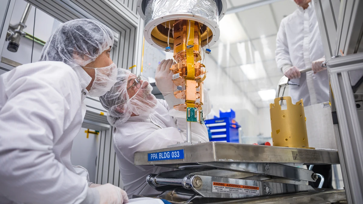
SQUIDs, or superconducting quantum interference devices, are the most sensitive magnetic sensors available. They detect signals from major organs, ranging from heart rhythms to brain activity, with sensitivity in the femtotesla range. Medical applications include magnetoencephalography (MEG), magnetocardiography (MCG), and liver iron concentration measurements. Coil-in-vacuum designs allow closer proximity to patients, improving resolution and reducing helium consumption. Advances in mechanical design, like adjustable tailpieces, help maintain sensor alignment despite thermal contraction.
Innovative cryostat designs using coil-in-vacuum setups have reduced helium boiloff and enabled clinical adoption of SQUID-based devices. Figure 5 shows a setup measuring magnetic susceptibility in the liver and an MEG system using a recondensing unit for zero boiloff operation.[8, 9]
Superconductivity is also revolutionizing radiation therapy. In 2015, the world’s first fully rotating superconducting gantry was installed at NIRS/HIMAC in Japan.[10] Using six combined-function magnets, it delivers carbon ion therapy with high precision. Its ability to rotate 360° allows targeted delivery without moving the patient, enabling Intensity Modulated Particle Therapy (IMPT).
New compact gantry designs use canted cosine theta magnets, allowing smaller, lighter systems with optimized beam optics.[11] The Next Ion Medical Machine Study (NIMMS) at CERN is building on this with designs for curved superconducting magnets and synchrotrons using NbTi and high temperature superconductors.[12] These developments aim to bring down costs and size, making advanced ion therapy more accessible.
Cryogenics and superconductivity have reshaped biomedical science over the past 50 years. Their applications, from precise imaging and diagnostics to life-saving therapies, highlight the value of continued investment and collaboration in these technologies.[13]
References
[1] Cooper, I., & Lee, A. (1961). Cryostatic congelation; a system for producing a limited controlled region of cooling or freezing of biological tissues. Journal of Nervous and Mental Disease, 133(4), 259–263.
[2] Dobak, J. (1998). A review of cryobiology and cryosurgery. Advances in Cryogenic Engineering, 43, 889–896.
[3] Baust, J. M., Robilitt, A., Gage, A., & Baust, J. G. (2016). Enhanced cryoablative methodologies. In Multiscale Technologies for Cryomedicine (Chapter 1). World Scientific Publishing.
[4] Schwall, R. E. (1987). MRI – Superconductivity in the marketplace. IEEE Transactions on Magnetics, 23(2), 1287–1293.
[5] Radebaugh, R., & Bar-Cohen, Y. (2016). Low temperature materials and mechanisms—Applications and challenges. In Low Temperature Materials and Mechanisms (Chapter 14). CRC Press.
[6] Boulant, N., Mauconduit, F., Gras, V., et al. (2024). In vivo imaging of the human brain with the Iseult 11.7-T MRI scanner. Nature Methods, 21, 2013–2016. https://doi.org/10.1038/s41592-024-02472-7
[7] Sarracanie, M., & Salameh, N. (2020). Low-field MRI: How low can we go? A fresh view on an old debate. Frontiers in Physics, 8, 172. https://doi.org/10.3389/fphy.2020.00172
[8] Brittenham, G. M., Farrell, D. E., Harris, J. W., Feldman, E. S., et al. (1982). Magnetic susceptibility of human iron stores. New England Journal of Medicine, 307(25), 1671–1675.
[9] Narasaki, K., et al. (2020). Development of zero boil-off cooling systems for superconducting self-shielded MEG. IOP Conference Series: Materials Science and Engineering, 755, 012107.
[10] Iwata, Y., et al. (2018). Superconducting gantry for carbon-ion radiotherapy. In Proceedings of the 9th International Particle Accelerator Conference (IPAC 2018). https://doi.org/10.18429/JACoW-IPAC2018-TUZGBF
[11] Benedetto, E., Harbi, N., et al. (2022). A carbon-ion superconducting gantry and a synchrotron based on canted cosine theta magnets. Physics in Medicine and Biology. https://doi.org/10.48550/arXiv.2105.04205
[12] Vretenar, M., et al. (2021). The next ion medical machine study at CERN: Towards a next generation cancer research and therapy facility with ion beams. In Proceedings of IPAC 2021. https://doi.org/10.18429/JACoW-IPAC2021-MOPAB413
[13] Rubinsky, B. (2000). Cryosurgery. Annual Review of Biomedical Engineering, 2, 157–187.